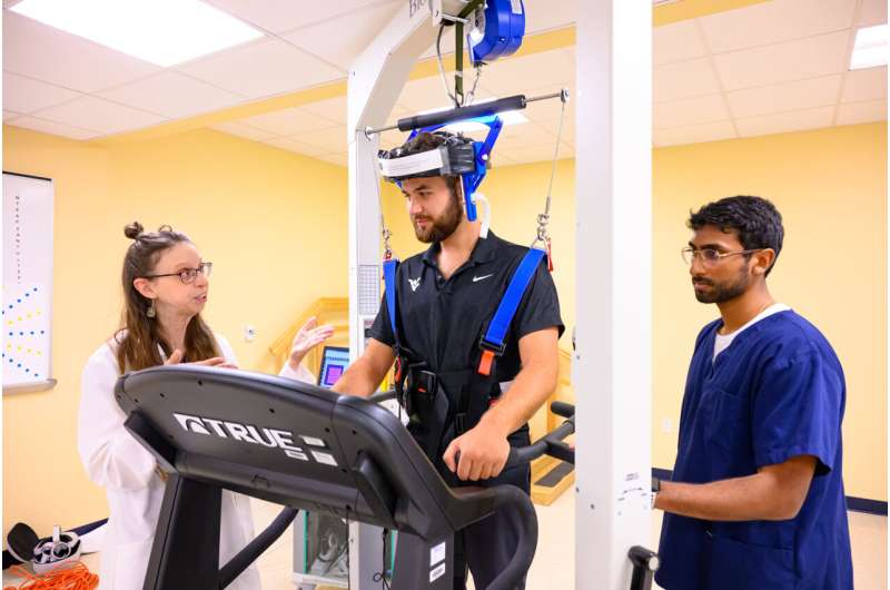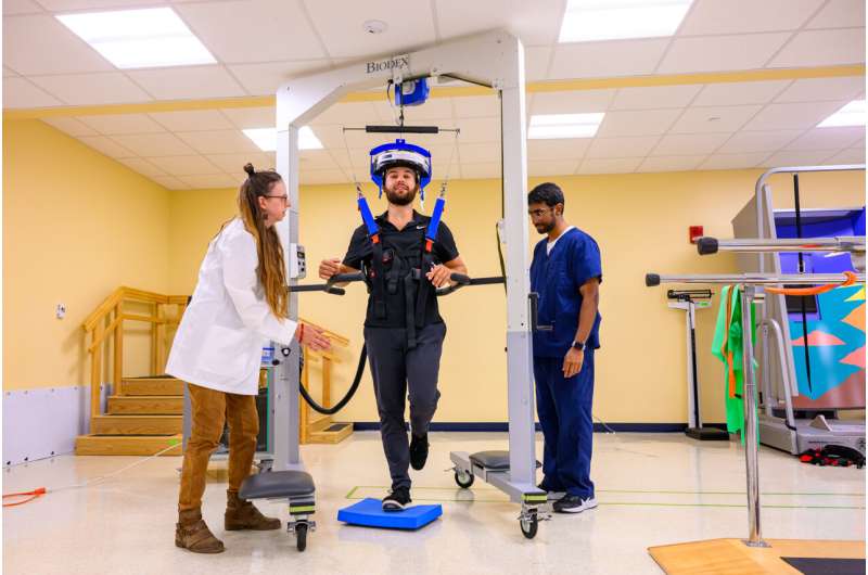
Julie Brefczynski-Lewis (left), a research assistant professor at WVU School Medicine, and students Colson Glover (center) and Nanda K. Siva demonstrate a walking PET scan at WVU Health Sciences. Credit: (WVU/Davidson Chan)
An upright neuroimaging device developed by West Virginia University neuroscientists, physicists and engineers that allows patients to move while undergoing a brain scan could help prioritize the development of imaging tools.
Researchers tested their prototype in a real-world environment to assess its accuracy and determine where improvements can be made. The prototype is being designed to address issues with traditional Positron Emission Tomography (PET) scanners, which require patients to lie still for imaging.
“PET imaging is widely used for research or diagnosis in patients with diseases that cause involuntary or uncontrollable movements, such as Parkinson’s disease. This makes it difficult or impossible to test these patients when their symptoms become too severe, because normal brain scans require you to remain very still.
“If you want to study human behavior, like walking, anxiety-provoking tasks, or even addiction, this device could also provide a way to capture images,” said Julie Brefczynski-Lewis, a research assistant professor in the Department of Neuroscience at the WVU School of Medicine and the Rockefeller Neuroscience Institute.
“It’s also useful for imaging patients with cognitive problems like dementia, because they have trouble holding still and even understanding the instructions they need to hold still, so they usually have to be sedated. If we wanted to image their brains while they were awake and alert, this would be a way to do that too.”
The scanner is an improved version of an earlier prototype that Brefczynski-Lewis and her team developed.
While both versions are hard hats, the first was heavier and allowed only slight side-to-side head movement. The new one, an Ambulatory Motion-enabling PET or AMPET, is lighter and fits the head like construction workers wear hard hats. A balanced support is located at the top.
“What we like about the AMPET is that it moves with the head, and you can be in a real environment where you’re immersed and you can walk with it,” she said. “What we showed in the study is that when the patients are walking, it doesn’t move relative to the head and that’s what allowed us to get a relatively clean image. We also wanted to see what could be improved by us or other labs that are making these devices.”
The studyconducted by Brefczynski-Lewis and colleagues from the School of Medicine, was published in the journal Communication medicine.
“The goal of this study was that we have this new technology and we can model it on a computer until the cows come home. But until you work with real patients, you don’t know how it’s really going to work in the real world,” she said.

WVU research assistant professor Julie Brefczynski-Lewis (left) and graduate students Colson Glover (center) and Nanda K. Siva hope their work on an upright neuroimaging device, pictured here, will expand the ability to study the brain in motion. Credit: (WVU/Davidson Chan)
To test the prototype, the team recruited volunteer outpatients who were scheduled for other scans and were already taking medications for imaging. Participants wearing the helmet walked around the scene while researchers looked at movement tolerance and assessed neural activity in motor brain areas.
“We observed brain activity in the parts of the brain that control the patients’ leg movements as they walked, which was exactly what we were hoping to see,” Brefczynski-Lewis said.
Their findings were further confirmed by observing a patient who had a prosthesis from hip to foot. His brain activity was shown mainly in the area that represented the natural leg.
“That was almost a separate test, which we didn’t expect,” she said.
To improve the prototype, researchers want to add a motion capture system and make the helmet larger so that a larger part of the brain can be imaged.
“Motion capture has been done for other technologies, so all we need to do is apply it to our device,” Brefczynski-Lewis said. “That will help because sometimes we miss the area of the brain that we want to see.”
Brefczynski-Lewis said the AMPET could be useful not only for imaging patients who can’t sit still, but also for neuroscientists studying human behavior and activities that involve natural movement, such as gestures, conversation and balance.
“To study balance, people are given MRI scans while lying down and are asked to imagine themselves being balanced. This is not the same as actually being balanced. A surface scan is also done, which does not show the deep brain structures,” she explains.
“You can see certain parts of the brain, but not the deeper parts of the brain that are typically more involved in things like movement, balance, emotion, memory, fear and joy.”
Brefczynski-Lewis and her team expect that in the future the AMPET will be used to monitor brain activity and treat people with PTSD, study mindfulness meditation, and integrate with virtual reality technologies.
“Being able to image the brain in motion shows that there is a whole new field that can open up with our device,” she said. “We can extend our neuroimaging research to human behavior that comes naturally: how we interact with the world and how we interact with others.
Along with Brefczynski-Lewis, the research was conducted by graduate students Nanda K. Siva of Parkersburg and Colson Glover of Lewisburg; Kaylee Nott of Point Pleasant, a West Virginia IDeA Network of Biomedical Research Excellence summer intern; Alexander Stolin, Gary Marano, Benjamin Parker, MaryBeth Mandich and James W. Lewis of West Virginia School of Medicine; Christopher Bauer, Sonia Chandi, Si Gao and Helen Melnick, alumni of the university; Stan Majewski, a former West Virginia researcher; and Jinyi Qi.
More information:
Nanda K. Siva et al, Real-time motion-assisted positron emission tomography of the brain of upright walking humans, Communication medicine (2024). DOI: 10.1038/s43856-024-00547-2
Quote: In practice, researchers develop a motion-compatible brain scanner (2024, August 7) Retrieved August 7, 2024 from https://medicalxpress.com/news/2024-08-motion-compatible-brain-scanner.html
This document is subject to copyright. Except for fair dealing for private study or research, no part may be reproduced without written permission. The contents are supplied for information purposes only.
 Healthy Famz Healthy Family News essential tips for a healthy family. Explore practical advice to keep your family happy and healthy.


